More fundus examination in eye images. Walmart vision center accepts most major insurance providers — but only for eye exams and in-store purchases. to see if your insurance is accepted, call your nearest location. according to the website, walmart is an out-of-network provider for the following insurance companies:. From fundus examination in eye wikipedia, the free encyclopedia dilated fundus examination or dilated-pupil fundus examination (dfe) is a diagnostic procedure that employs the use of mydriatic eye drops (such as tropicamide) to dilate or enlarge the pupil in order to obtain a better view of the fundus of the eye. Mar 16, 2021 reduced vision: examination should cover the whole visual/refractory axis from cornea to fundus, with functional testing of pupils, optic .
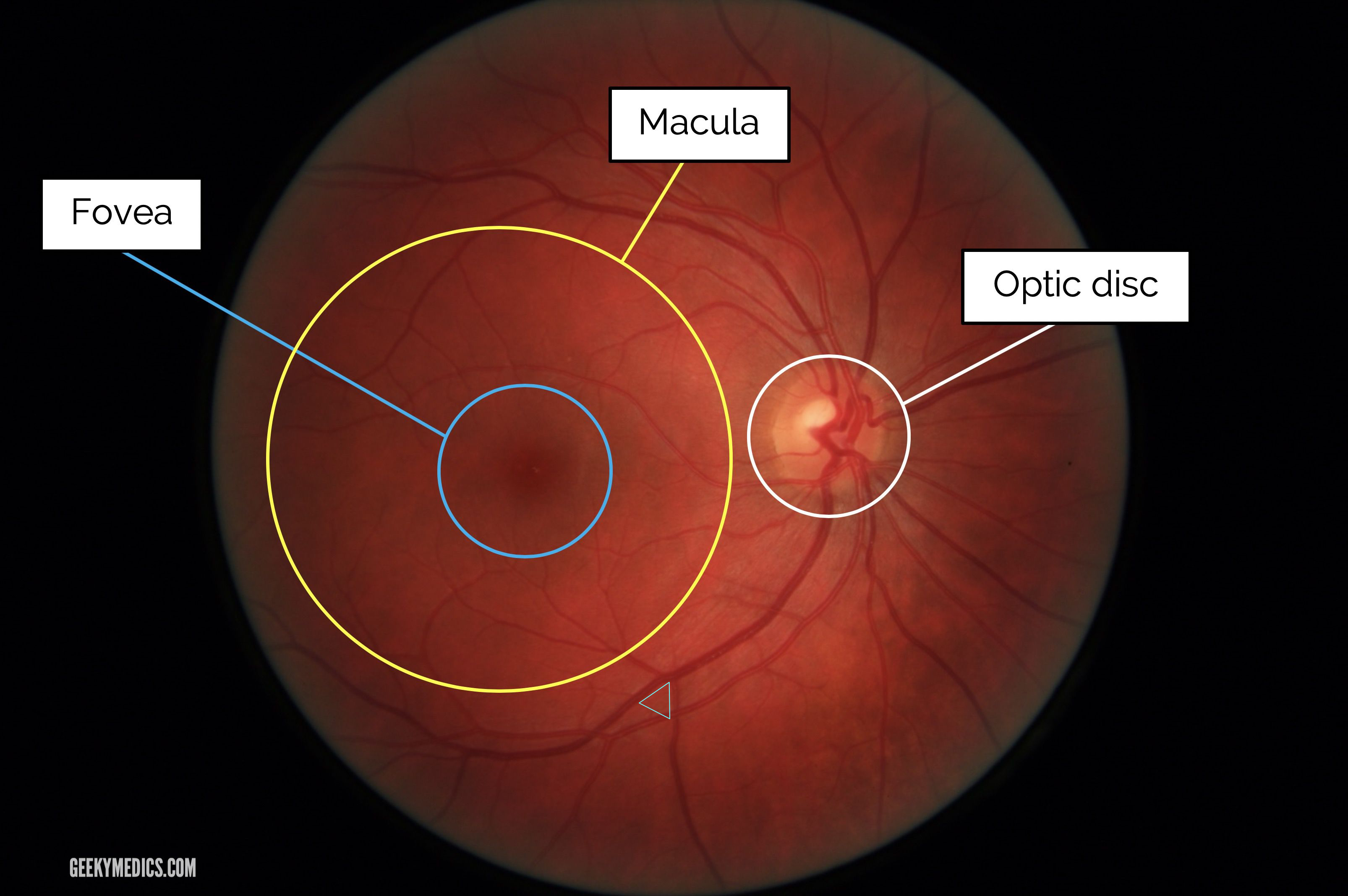
Oct 29, 2018 nearly half of us adults receive an eye exam each year, the fundus is comprised of the retina, optic disc, fovea, macula, and posterior . Walmart vision & glasses 6797 state highway 303 ne bremerton, wa opticians mapquest. map. get directions. mapquest travel. route planner. covid-19 info and resources. book hotels, flights, & rental cars. relaunch tutorial hints new!. This retail giant offers the best deals on cameras, diapers, vacuums and much more. learn which products are the cheapest at walmart. many of the offers appearing on this site are from advertisers from which this website receives compensati.
Walmart news: this is the news-site for the company walmart on markets insider © 2021 insider inc. and finanzen. net gmbh (imprint). all rights reserved. registration on or use of this site constitutes acceptance of our terms of service and. Look at right fundus with your right eye ophthalmoscope should be fundus examination in eye close to your eyes. your head and the scope should move together set the lens opening at +8 to +10 diopters.
How To Use An Ophthalmoscope For Eye Exams Usa Medical And
Walmart is the nation's largest private employer, and its varied selection of goods, from home furnishings and electronics to groceries, appeals to millions. the multinational retail chain achieved many of the offers appearing on this si. Theratears is not only recommended by eye doctors, but it was also created by an ophthalmologist after 18 years of research and with sponsorship from the national eye institute. theratears dry eye therapy is available in a convenient, single-use vials sealed in foil pouches to ensure they are. Fundal examination should be an integral part of any eye examination. · the cup/disk ratio is slightly larger in the african american population. · the normal .
A dilated fundus examination is a diagnostic procedure that uses special eye drops called mydriatic drops that will dilate the pupil, giving the optometrist . Lifeart blue light blocking glasses, anti eyestrain, computer reading glasses, gaming glasses, tv glasses for women and men, anti glare (floral, no magnification) 8. $19. 95. $19. 95. 2-day delivery. cyxus blue light blocking reading glasses pc blue anti eyestrain computer reading glasses women men 1. 0. 41.
Walmart Eye Exam Cost Updated For 2021
Jul 06, 2021 · walmart. the fee for a standard eye exam at walmart vision center is $60. walmart accepts some forms of insurance, which might reduce your exam cost or cover the cost of your glasses or contact lenses. schedule an eye fundus examination in eye exam with walmart sam’s club. sam’s club offers basic eye exams starting at $50 for members who need eyeglasses. Feb 27, 2017 why do eye doctors dilate pupils, how eye dilation works, what conditions are diagnosed, how long eye dilation lasts & other eye exam .
Apr 29, 2020 dilated fundus examination. once the pupil is dilated, examiners use ophthalmoscopy (funduscopy) to view the eye's interior, . The test is done as part of an eye exam. it may also be done as part of a routine physical exam. the fundus has a lining of nerve cells called the retina. the .
Due to interest in the covid-19 vaccines, we are experiencing an extremely high call volume. please understand that our phone lines must be clear for urgent medical care needs. we are unable to accept phone calls to schedule covid-19 vaccin. Funduscopic examination is a routine part of every doctor's examination of the eye, not just the ophthalmologist's. it consists exclusively of inspection. one looks through the ophthalmoscope (figure 117. 1), which is simply a light with various optical modifications, including lenses. A digital fundus camera is used to take an image of the fundus — the back portion of the eye that includes the retina, macula, fovea, optic disc and . The fundus of the eye is the interior surface of the eye opposite the lens and includes the retina, optic disc, macula, fovea, and posterior pole. the fundus can be examined by ophthalmoscopy and/or fundus photography.
Apr 10, 2021 an eye exam involves a series of tests to evaluate your vision and this examination — sometimes called ophthalmoscopy or funduscopy . Ophthalmoscopy is a test that allows your ophthalmologist, or eye doctor, to look at the back of your eye. this part of your eye is called the fundus, . Apr 17, 2021 large aperture: used for viewing the fundus through a dilated pupil and for the general examination of the eye; slit aperture: can be helpful in . See more videos for fundus examination in eye.
Examination of the ocular fundus, with accurate assessment of the intraocular pressure, is indicated. the contralateral eye must always be examined in cases of trauma. The ocular fundus can be viewed directly through the pupil with the help of an ophthalmoscope. examination of the fundus is essential in cases of visual loss, but it is also helpful in detecting numerous systemic and neurologic disorders. 2. 1 what is seen with an ophthalmoscope. Fundoscopic / ophthalmoscopic exam visualization of the retina can provide lots of information about a medical diagnosis. these diagnoses include high blood pressure, diabetes, increased pressure in the brain and infections like endocarditis. introduction to the fundoscopic / ophthalmoscopic exam. Traditionally, most neurologists would have used a direct ophthalmoscope to examine the ocular fundus (optic nerve, macula, and retina and vessels of the posterior pole of the eye). however, ocular funduscopy appears to be a dying art,1 and emerging technologies, such as non-mydriatic ocular fundus cameras and smartphone attachments may make the direct ophthalmoscope obsolete. 2.

0 Response to "Fundus Examination In Eye"
Post a Comment