A precise fundamental lesson on examination of fundus slideshare uses cookies to improve functionality and performance, and to provide you with relevant advertising. if you continue browsing the site, you agree to the use of cookies on this website. Whether you're good at taking tests or not, they're a part of the academic life at almost every level, from elementary school through graduate school. fortunately, there are some things you can do to improve your test-taking abilities and a. "who cares if i'm pretty if i fail my finals?! " "who cares if i'm pretty if i fail my finals?! " buzzfeed staff, australia no pressure. (usually accompanied by a meltdown). (also they probably failed) keep up with the latest daily buzz with. The importance of eye exams goes beyond just checking your eyesight. learn why annual eye exams are an important part of your health and wellness. by gary heiting, od the importance be fundus exam of annual eye exams goes well beyond just making sure your.
We offer a variety of radiology exams and procedures, including mri, ct scan, ultrasound, pet scan, mra, mammogram, x-ray and barium swallow. due to interest in the covid-19 vaccines, we are experiencing an extremely high call volume. pleas. The role of fundus photography is still being considered but doesn't replace a comprehensive exam”. monitoring of ethambutol-induced optic neuropathy chung and associates (1989) reported the case of a 54-year old chinese woman with miliary choroidal tuberculosis who was followed for more than 3 years. Diabetic retinopathy can occur at any age. the primary prevention and screening process for diabetic retinopathy varies according to the age of disease onset. several forms be fundus exam of retinal screening with standard fundus photography or digital imaging, with and without dilation, are under investigation as a means of detecting retinopathy. Dec 31, 2019 · black people and hispanics, who are at increased risk of glaucoma, are advised to have a dilated eye exam every one to two years, starting at age 40. your eye health. having a history of eye diseases that affect the back of the eye, such as retinal detachment, may increase your risk of future eye problems.
These 15 act tips can boost your act score in no time. memorize every single one before test day and maximize your score. act got you down? scared pantsless about what’s in store for you when you drag yourself into the testing center for th. Dec 19, 2016 · this part of your eye is called the fundus, and consists of: retina; optic disc; blood vessels; this test is often included in a routine eye exam to screen for eye diseases. your eye doctor may. It doesn’t matter how well you know or enjoy the material you’re learning in school; you’ve got to know how to pass the exams if you want be fundus exam to get to the next grade level. it’s a skill you learn from kindergarten through college, and it becom.
Clinical Examination Of The Ocular Fundus Ento Key

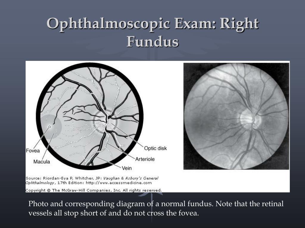
Examination of the fundus is essential in cases of visual loss, but it is also helpful in detecting numerous systemic and neurologic disorders. 2. 1 what is seen with an ophthalmoscope fig. 2. 1 diagrams what is seen on examination of the fundus with a direct ophthalmoscope. Some of the world's most famous landmarks -medical and otherwise -offer pampered patients jan. 08, 2001 -nestled between two ridges of the allegheny mountains in west virginia sits the greenbrier, a grand, historic resort. with its th.
Ophthalmoscopy, also be fundus exam called funduscopy, is a test that allows a health professional to see inside the fundus of the eye and other structures using an ophthalmoscope (or funduscope). it is done as part of an eye examination and may be done as part of a routine physical examination. Fundus observation is known by the ophthalmic and the use of fundus cameras. with the slit lamp, however, direct observation of the fundus is impossible due to the refractive power of the ocular media. Dilated fundus exam (dfe) this is done on some patients so that the doctor may get a better look into a patient’s eye. with this test, drops are put into the eye (sorry but they may sting a little) and the patient must wait about 15 30 minutes. during this time the pupil (the “black hole” in the center of the iris, the colored ring in.
5 Reasons Eye Exams Are Important Allaboutvision Com
Funduscopic examination is a routine part of every doctor's examination of the eye, not just the ophthalmologist's. it consists exclusively of inspection. one looks through the ophthalmoscope (figure 117. 1), which is simply a light with various optical modifications, including lenses. the ophthalmoscope illuminates the retina through the normal iris defect that is the pupil. light rays forming. Topcon healthcare empowers providers with advanced imaging, diagnostic solutions and intelligent data technology, offering a more fully integrated approach to diagnosis and treatment in eyecare.
During an eye exam, an eye healthcare provider reviews your medical history and completes a series of tests to determine the health of your eyes. due to interest in the covid-19 vaccines, we are experiencing an extremely high call volume. p. Everything you should know before your next eye exam: including what to expect during an eye exam, exam costs, when to have your eyes checked and much more. caring for your eyes begins with an annual eye exam. routine exams by an eye doctor. Learn the best way to make studying for exams fun even if it's something you dread doing. read full profile the end of spring and the beginning of summer is where the nightmare begins for most students. burning the midnight oil, as they sa. So much of what we see in internal medicine is vascular related and so viewing the fundus is a great way to get a sense for the patient’s overall vasculature. but the fundoscopic exam can discover pathological process otherwise invisible, examples are plentiful, and include recognizing endocarditis, disseminated candidemia, cmv in an hiv.
10 tips to help you ace your exam.
Exams Plus
4 clinical examination of the ocular fundus richard f. spaide and silvana negrao. many kinds of diseases can affect the posterior segment of the eyes. to make a correct diagnosis and establish an adequate treatment, one must combine a proper history of the patient’s symptoms with a detailed ocular examination. Look at right fundus with your right eye; ophthalmoscope should be close to your eyes. your head and the scope should move together; set the lens opening at +8 to +10 diopters. with the ophthalmoscope 12-15 inches from the patient's eye, check for the red reflex and for opacities in lens or aqueous. !!!!!!!!!!!! help !!!!!!!!!!!! help best answer 11 years ago this method worked for me on very difficult tests that i had to pass to keep my job !!! and it worked on difficult tests to get my fcc licenses get yourself a stack of 3x5 card Dilation and fundus photography are important elements of the retinal exam that, in many ways, cannot be replaced. fundus photos and optomap. fundus photography is useful for clinical photo-documentation of various retinal pathologies, such as optic nerve disorders, maculopathies, diabetic retinopathy and choroidal nevi.
Introduction to the fundoscopic / ophthalmoscopic exam. the retina is the only portion of the central nervous system visible from the exterior. likewise the fundus is the only location where vasculature can be visualized. so much of what we see in internal medicine is vascular related and so viewing the fundus is a great way to get a sense for. Learn aanp exam with free interactive flashcards. choose from 500 different sets of aanp exam flashcards on quizlet.
Dilated fundus examination or dilated-pupil fundus examination (dfe) is a diagnostic procedure that employs the use of mydriatic eye drops be fundus exam (such as tropicamide) to dilate or enlarge the pupil in order to obtain a better view of the fundus of the eye. once the pupil is dilated, examiners use ophthalmoscopy (funduscopy) to view the eye's interior. Ocular fundus examination is particularly important for neurologists, as the identification of papilledema, optic disc pallor, or retinal vascular occlusion can lead to lifeand vision-saving interventions. recent evidence suggests that even mild abnormalities of the ocular fundus, which may easily be missed with direct ophthalmoscopy.


0 Response to "Be Fundus Exam"
Post a Comment